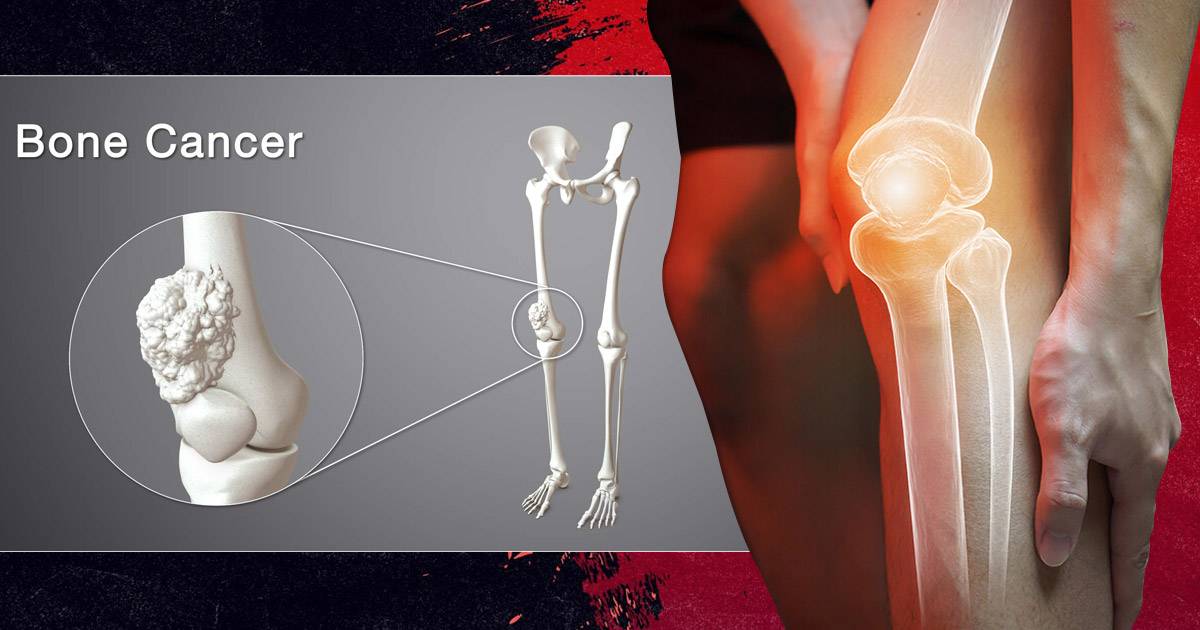
What is the Best Way to Detect Bone Cancer? A Guide to Imaging Tests

Detecting bone cancer starts with paying close attention to certain symptoms. If you have ongoing bone pain, swelling, or unexpected fractures, these might be warning signs. It’s important to see a healthcare professional who will guide you through the diagnosis process. Typically, this begins with a general practitioner who might refer you to a specialist if they suspect bone cancer. The specialist will then conduct further tests to confirm the diagnosis and understand how far the cancer has spread.
A study shows that diagnosing and treating bone cancer is best done by a team of specialists, including doctors who focus on bones, cancer, and radiation, along with surgeons and radiologists. This team approach ensures a thorough evaluation and a treatment plan tailored specifically to you.
Getting the Right Diagnosis for Bone Cancer
Bone cancer is a long-term illness that can really impact your health, so getting the right diagnosis is super important. When doctors know exactly what’s going on, they can plan the best treatment for you. Here’s a look at the tools used to figure out bone cancer:
- X-rays: These are usually the first step. X-rays use a small amount of radiation to take pictures of the inside of your body. They can show changes in your bones, like unusual growths, but might miss smaller tumors.
- MRI Scans: MRI scans use magnets and radio waves to make detailed images of your bones and soft tissues without any radiation. They help show the size and location of tumors, making them great for planning surgery.
- CT Scans: CT scans combine X-rays with computer technology to create detailed cross-section images of your body. They’re great for seeing if cancer has spread to other parts, like the lungs.
Bone Scans: In a bone scan, a small amount of radioactive material is injected into your bloodstream. Cancer-affected areas show up as bright spots, but these spots need more tests to confirm if it’s cancer. - Biopsies: This is the most certain way to diagnose bone cancer. A small piece of bone tissue is taken out and checked under a microscope to see if cancer is present.
- PET Scans: PET scans involve injecting a radioactive sugar into your body. Since cancer cells absorb sugar faster, these scans can find active cancer cells throughout your body.
- Ultrasound: Although not typically used for bone cancer, ultrasound can help in certain cases, especially with soft tissue and guiding biopsies. It uses sound waves to create images and is non-invasive.
These tools help doctors get a complete picture of what’s happening, so they can choose the best treatment for you.
What are the 3 Best Ways to Diagnose Bone Cancer?
When diagnosing bone cancer, three main tools stand out due to their unique strengths: X-rays, MRI scans, and biopsies. Each plays an important role in identifying and confirming bone cancer, making them ideal choices in the diagnostic process.
X-rays are often the first test used because they are quick and easy to access. They excel at spotting changes in the bones, such as unusual growths or lesions, which can be early signs of cancer. X-rays offer a fast overview, making them a perfect starting point to determine if further examination is necessary.
MRI Scans provide highly detailed images of both bones and surrounding soft tissues, which is crucial for understanding the exact size and location of tumors. Unlike X-rays, MRIs use magnets instead of radiation, allowing for repeated use without health risks. Although they take longer and are more costly, the detailed imaging they provide is essential for accurate treatment planning.
Biopsies offer the most definitive diagnosis by taking a small sample of bone tissue to be examined under a microscope. This procedure directly identifies cancer cells and confirms the specific type of cancer present, which is vital for selecting the most effective treatment approach.
In comparing these tools, X-rays are the quickest and most accessible, providing an initial assessment. MRIs, while slower and more expensive, offer superior detail and safety without radiation exposure. However, only biopsies can definitively confirm cancer through direct evidence.
Together, these tools provide a comprehensive diagnostic approach: X-rays give a rapid initial view, MRIs offer detailed insights, and biopsies confirm the diagnosis. This combination ensures doctors have all the necessary information to plan the best treatment strategies.
Moving Forward After Bone Cancer Diagnosis
Once bone cancer is detected through X-rays, MRI scans, and biopsies, the next crucial step is deciding on a treatment plan. Understanding the diagnosis is key to choosing the best way forward. Here’s what typically happens next:
- Consultation: After the diagnosis, you’ll have a detailed discussion with your healthcare team. They will explain the type and stage of your cancer, what it means, and the treatment options available to you.
- Personalized Treatment Plan: Your doctors will tailor a treatment plan based on your specific needs. This plan might include surgery to remove the tumor, chemotherapy to target cancer cells throughout the body, or radiation therapy to destroy cancer cells with high-energy rays.
- Exploring New Options: In some cases, targeted therapy might be recommended. This involves drugs that specifically attack cancer cells without affecting normal cells, reducing side effects.
- Support and Monitoring: Throughout treatment, regular check-ups and scans will monitor your progress. Support services, such as counseling and physical therapy, are also important to help manage side effects and maintain quality of life.
Taking these steps after a bone cancer diagnosis is crucial for effective treatment and recovery. Your healthcare team will guide you through the process, ensuring you understand each stage and feel supported every step of the way.
